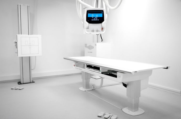
Digital X-Ray System - Sinogram
Further information
During a sinogram, a contrast dye is injected into the ureters through a small catheter that is inserted into the urethra (the tube that carries urine out of the bladder). The contrast dye helps to highlight the ureters and makes them visible on X-ray images.
After the contrast dye is injected, a series of X-rays are taken as the dye moves through the ureters and into the bladder. This allows the radiologist to evaluate the structure of the urinary tract and look for any abnormalities, such as blockages or strictures.
Sinograms were once a common diagnostic tool for evaluating the urinary tract, but they have largely been replaced by newer imaging technologies such as computed tomography (CT) and magnetic resonance imaging (MRI), which provide more detailed images without the need for invasive procedures. However, sinograms may still be used in some situations, particularly when other imaging tests are inconclusive or unavailable.
